What is skull base surgery?
The skull base is the bony floor on which the brain rests. It sits behind the eyes and nose and above the ears and neck. Many important nerves pass through it, including the nerves for sight, smell, hearing, facial movement and swallowing, along with major blood vessels.
Read moreShow less
Tumours in this area include meningiomas, acoustic neuromas (also called vestibular schwannomas, growths on the hearing and balance nerve), pituitary tumours, craniopharyngiomas and chordomas. Most are not cancer, but they can cause problems by pressing on nerves or the brain.
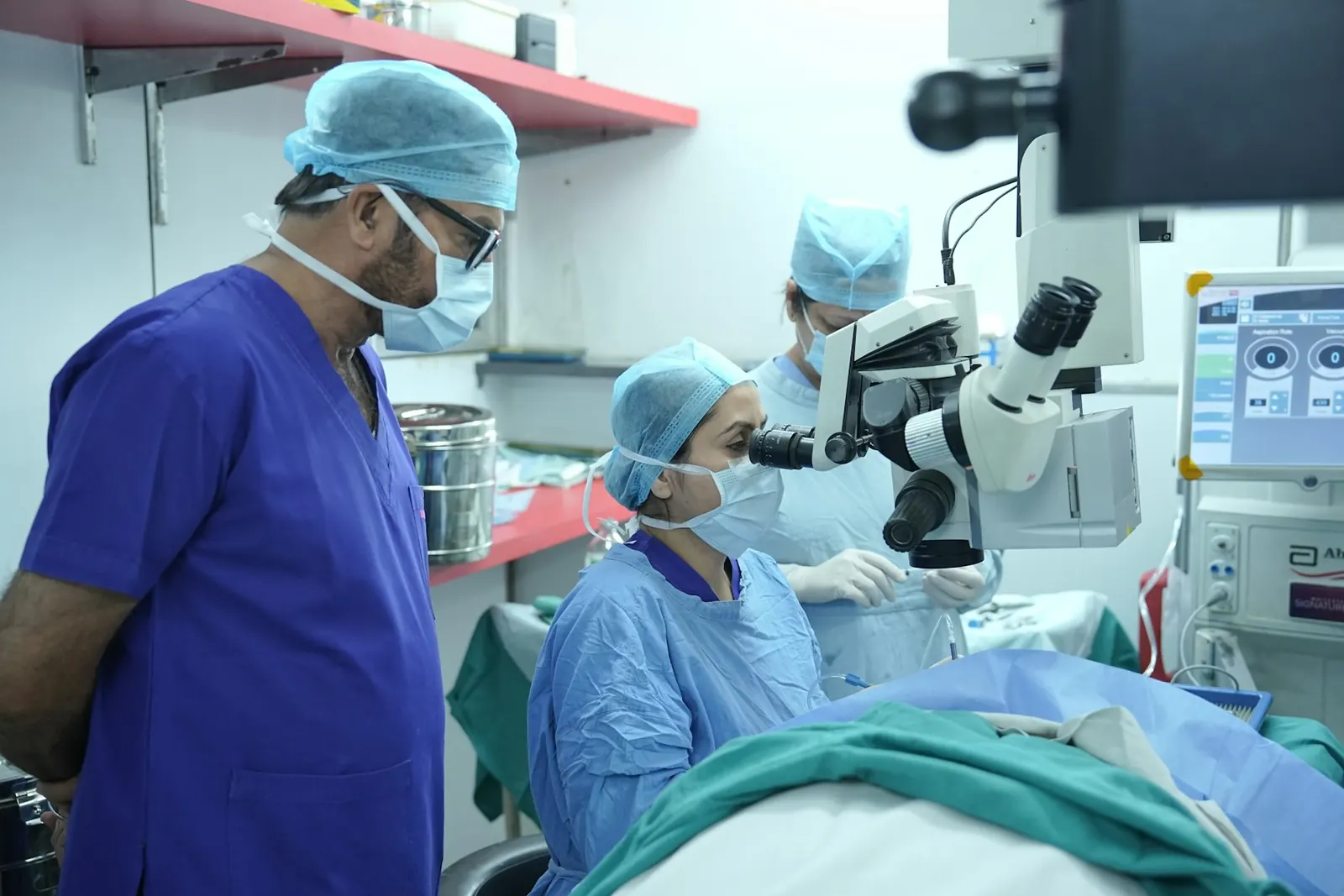
Depending on where the tumour is, the surgeon may reach it through the nose using an endoscope (endoscopic endonasal approach), or through an opening in the skull using a microscope (microscopic approach). Some operations combine both, and an ENT (ear, nose and throat) surgeon may be part of the team.
Who may need it
Surgery is one option, not the only one. It is decided after an examination and a careful look at your scans, and non-surgical treatment is discussed first where it is suitable.
How it is done
- 1
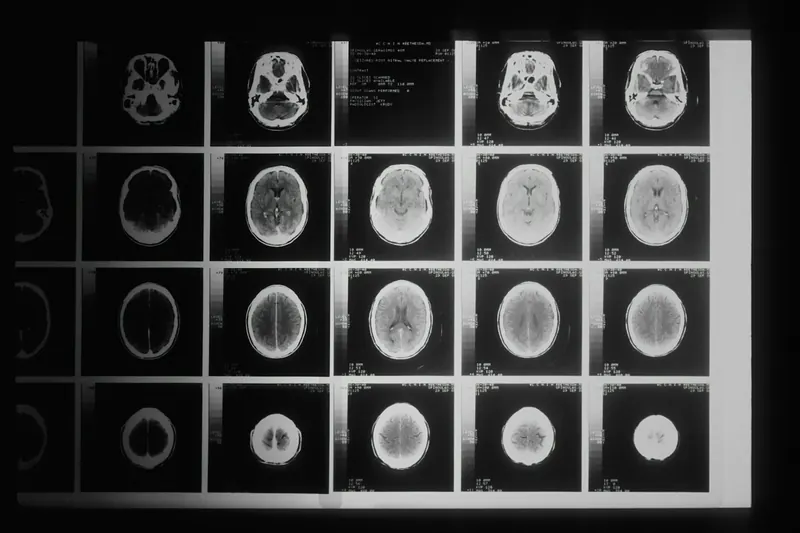
Detailed MRI and CT scans are studied to plan the safest route. Some patients need eye, hearing or hormone tests first.
- 2
You are given general anaesthesia and are fully asleep.
- 3
In the endoscopic approach, the surgeon passes a thin camera and tools through the nostrils to reach the tumour, with no cut on the face. In the microscopic approach, a small opening is made in the skull, often behind the ear or near the hairline.
- 4
The tumour is carefully separated from nerves and blood vessels and removed as far as is safe. Sometimes a part is left on purpose to protect important structures, and treated later if needed.
- 5
The opening in the skull base is repaired, sometimes using a flap of tissue from inside the nose or a small graft, to prevent a CSF leak.
- 6
You are watched closely in the ICU (intensive care unit) after the operation.
Possible risks
Every operation carries some risk. Your surgeon will explain which of these apply to you.
Preparing for surgery
Recovery
After surgery, patients usually spend a day or more in the ICU. The total hospital stay is often about 5 to 10 days, and sometimes longer, depending on the operation and recovery.
Read moreShow less
After surgery through the nose, avoid blowing your nose, straining, lifting heavy weights and bending forward for some weeks, as advised. Report any clear watery drip from the nose, severe headache or fever at once.
Some nerve problems improve slowly over months. Physiotherapy, speech and swallowing therapy, or eye care may be needed. Regular MRI scans are usually advised to check for any regrowth.
Questions patients ask
Will there be a scar on my face?
When the surgery is done through the nose with an endoscope, there is no cut on the face. When an opening in the skull is needed, the cut is usually placed behind the ear or within the hairline.
Is endoscopic surgery always better than open surgery for the skull base?
No. Each approach suits different tumour positions. Some tumours are easier and safer to reach through the nose, others through the skull, and some need both. The surgeon chooses the route that gives the safest access to your tumour.
Can I avoid surgery?
Sometimes. Small tumours that are not growing or causing symptoms may be watched with MRI scans. Some can be treated with focused radiation. Your surgeon will explain whether these options suit you.
How long will I stay in hospital?
Often about 5 to 10 days, including a stay in the ICU. It may be longer for larger operations or if there are complications.
Will my hearing or vision come back after surgery?
It depends on how long and how badly the nerve was pressed before surgery. Vision often improves when pressure on the optic nerve is relieved early, but hearing that is already lost often does not return. Your surgeon will discuss what to expect in your case.
Disclaimer: This information is for general awareness and is not a substitute for a medical consultation. Read moreSource: Content on this website was prepared with the help of AI tools (AI-assisted) and is medically reviewed by Dr. Kshitiz Nigam, MBBS, MS, MCh (Neurosurgery), Reg. No. MP-33282.